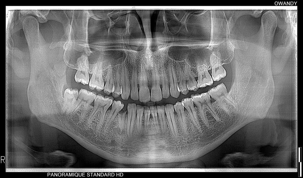
UNRIVALLED IMAGE QUALITY
New-generation dental panoramic
Available in 2D and 3D versions


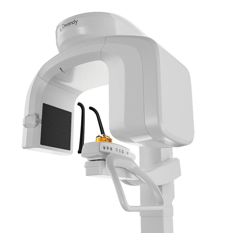
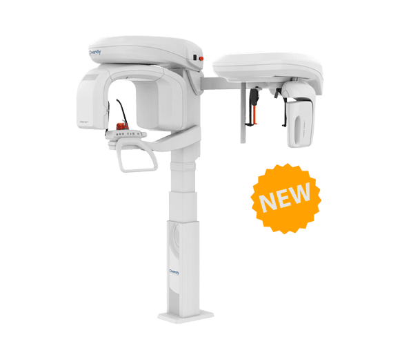
I-Max 3D XPRO
New smart Cone Beam generation
Embrace dental diagnostics future's and improve your daily practice.
Ask more to your panoramic
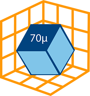
Technologie Super IGZO
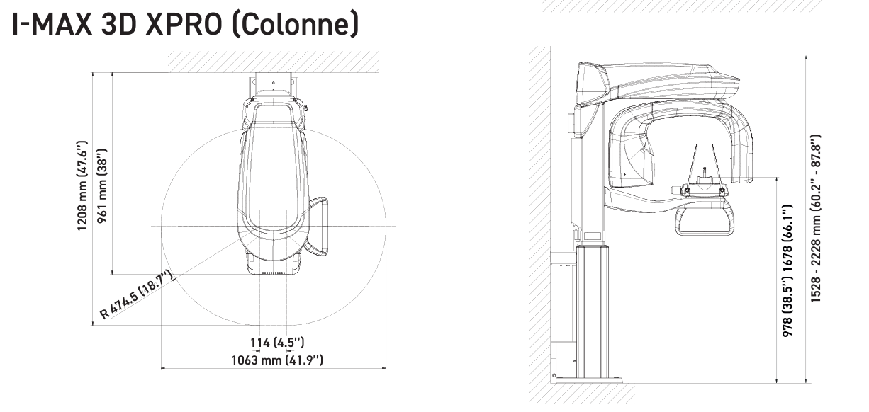
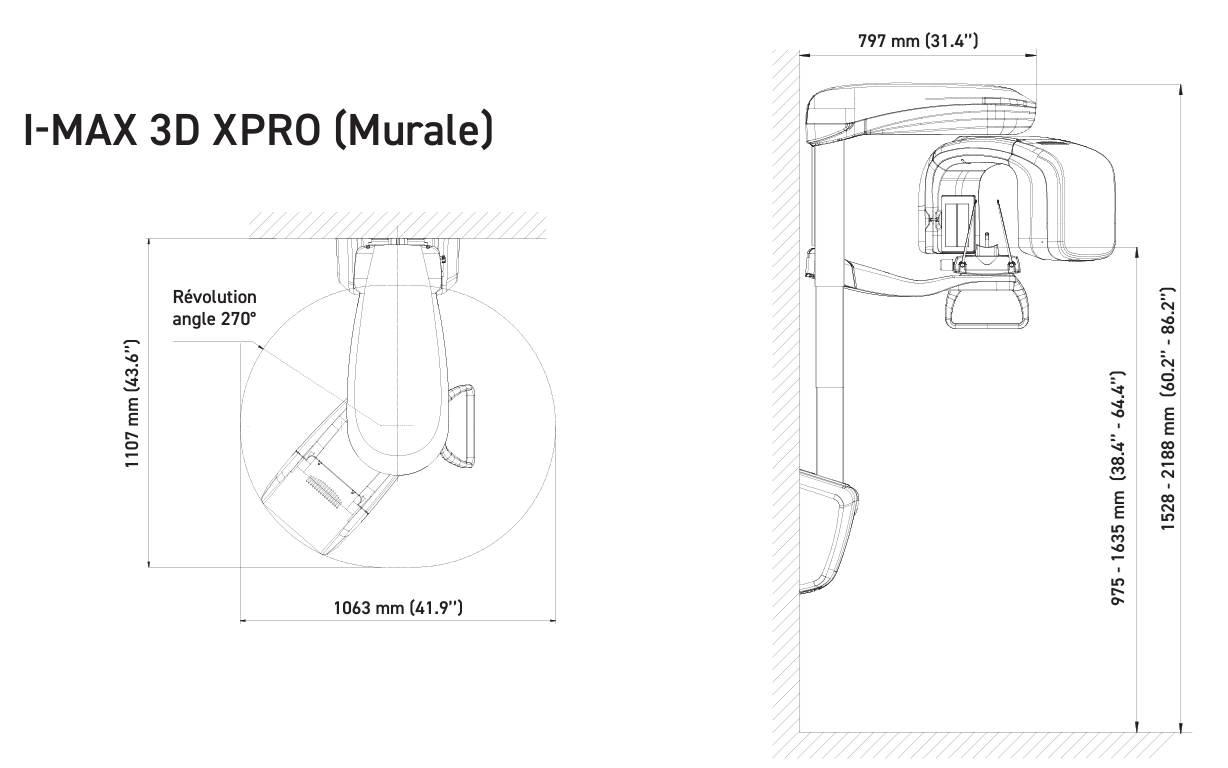
I-Max 3D XPRO adapts to all practices
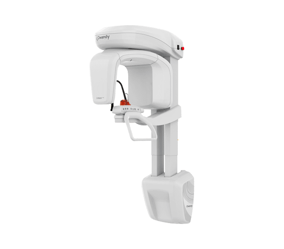
Available in 2 versions with different attachment options.
Your dental panoramic adapts to the different configurations of your dental practices.
I-MAX 3D XPRO version (Column)
I-MAX 3D XPRO version (wall-mounted)
L’I-Max 3D XPRO
CONE BEAM MULTI-FOV ( Field of view)
16 x 11 CM
Full dentition, TMJ, sinus, airways (Optional)

12 x 10 CM
Full dentition with condyles

9 x 9 CM
Full dentition

9 x 5 CM
Full arch
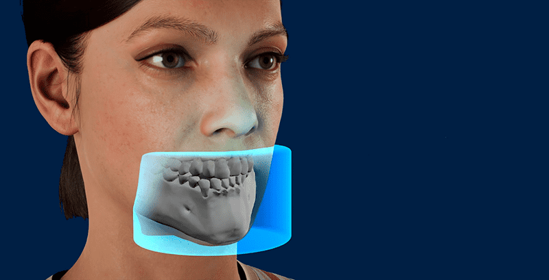
5 x 5 CM
Localized volume
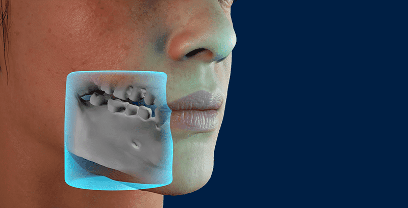
Although there are 1001 ways to treat a case, there is only one correct diagnosis.
I-Max 3D XPRO is a 4-in-1 dental Cone Beam designed to enable more accurate diagnosis, treatment planning and better patient outcomes, our dental panoramic integrates seamlessly into the digital workflow.
From imaging to guided surgery, it guarantees the highest levels of precision and safety.
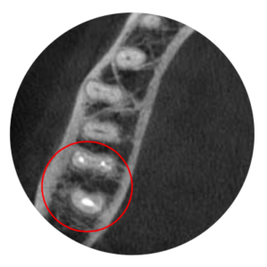
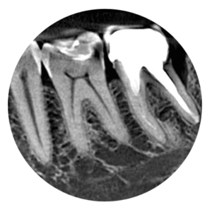
AutoMAR (Automatic Metal Artefact Reduction)
Rayonnement réduit des artefacts grâce à notre nouvel algorithme.
without autoMAR
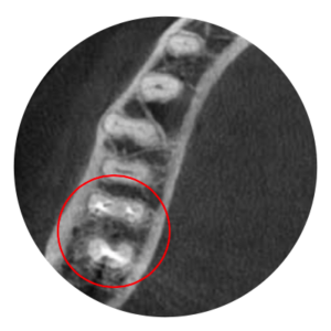
with autoMAR
side view

Filter and processing algorithm
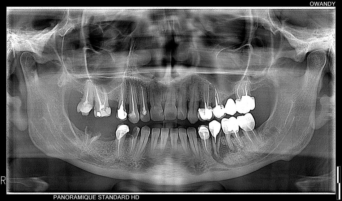
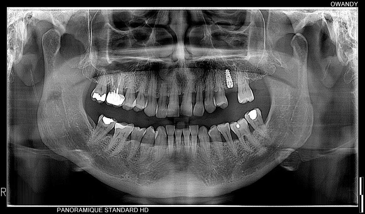
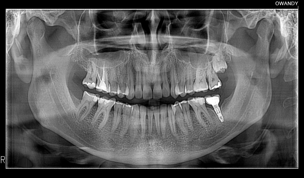
Thanks to Super IGZO technology, our new generation of intelligent sensors delivers exceptional image quality.
Move the cursor to compare CMOS sensor radio quality versus a Super IGZO sensor.
I-Max 3D XPRO, intuitive and easy to use
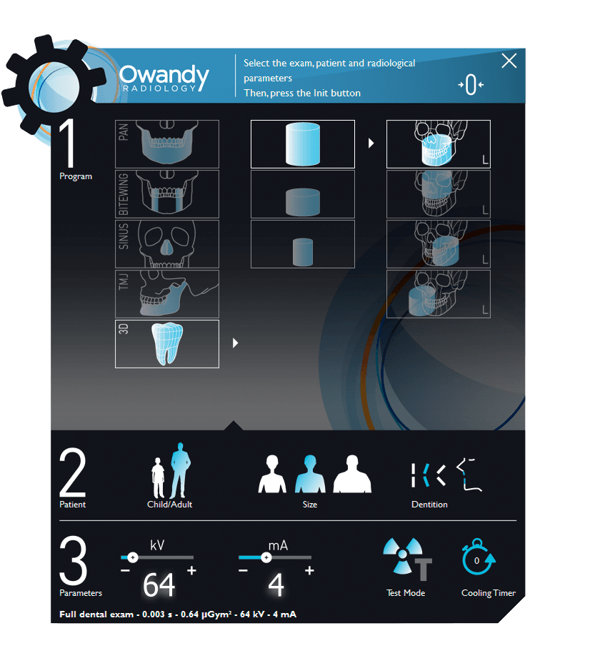
The I-Max 3D XPRO panoramic unit adapts to you and your current needs. It allows you to switch from conventional 2D to a 2D/3D Cone Beam version whenever you want.
Thanks to its intuitive interface, you can navigate easily between 2D and 3D examinations.
See all our demo videos on our website and our Owandy Radiology YouTube channel.
Discover it in just a few minutes
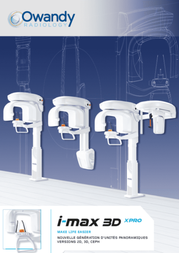
I-Max 3D XPRO Brochure
Find out more about the I-Max 3D XPRO in a video and in our documents.
Discover the latest innovations integrated into our XPRO panoramic range
Latest SUPER IGZO technology generation.
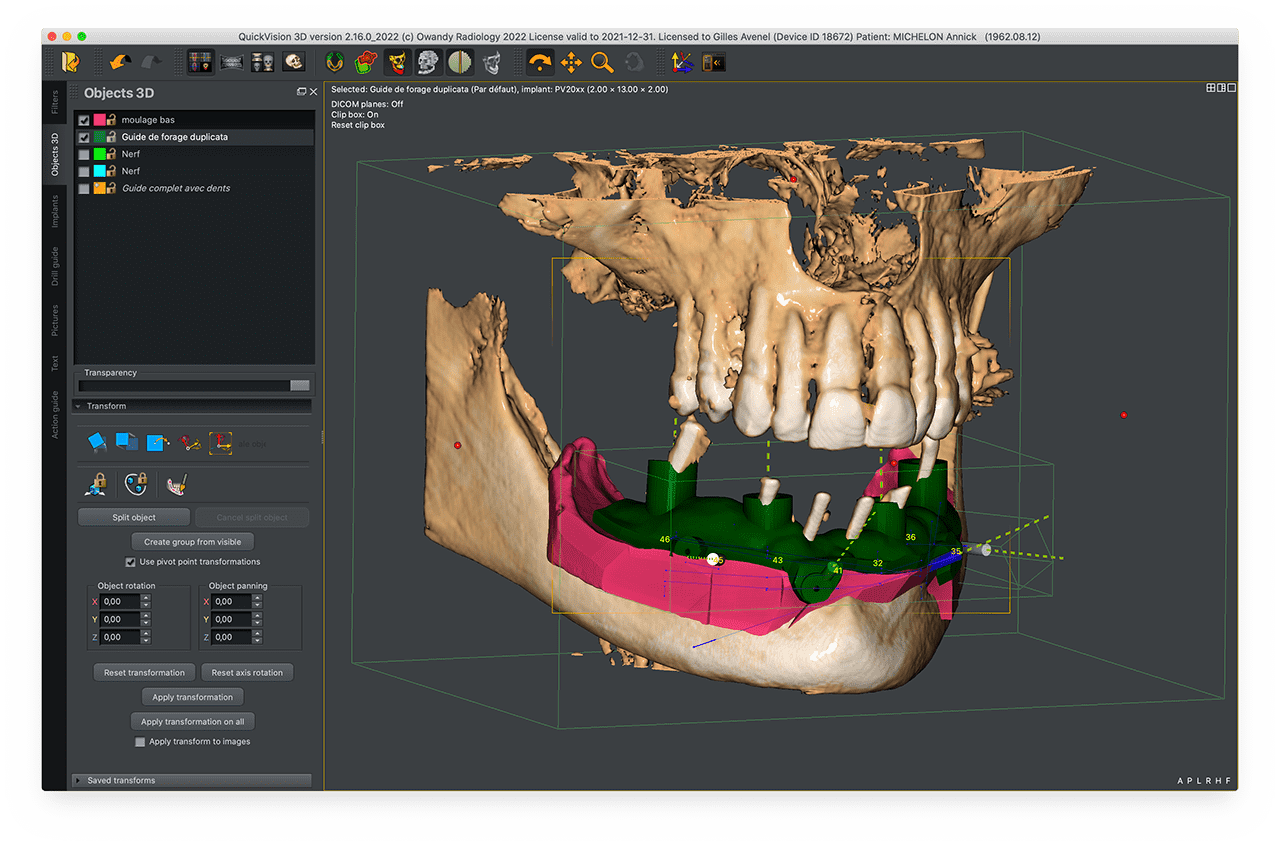
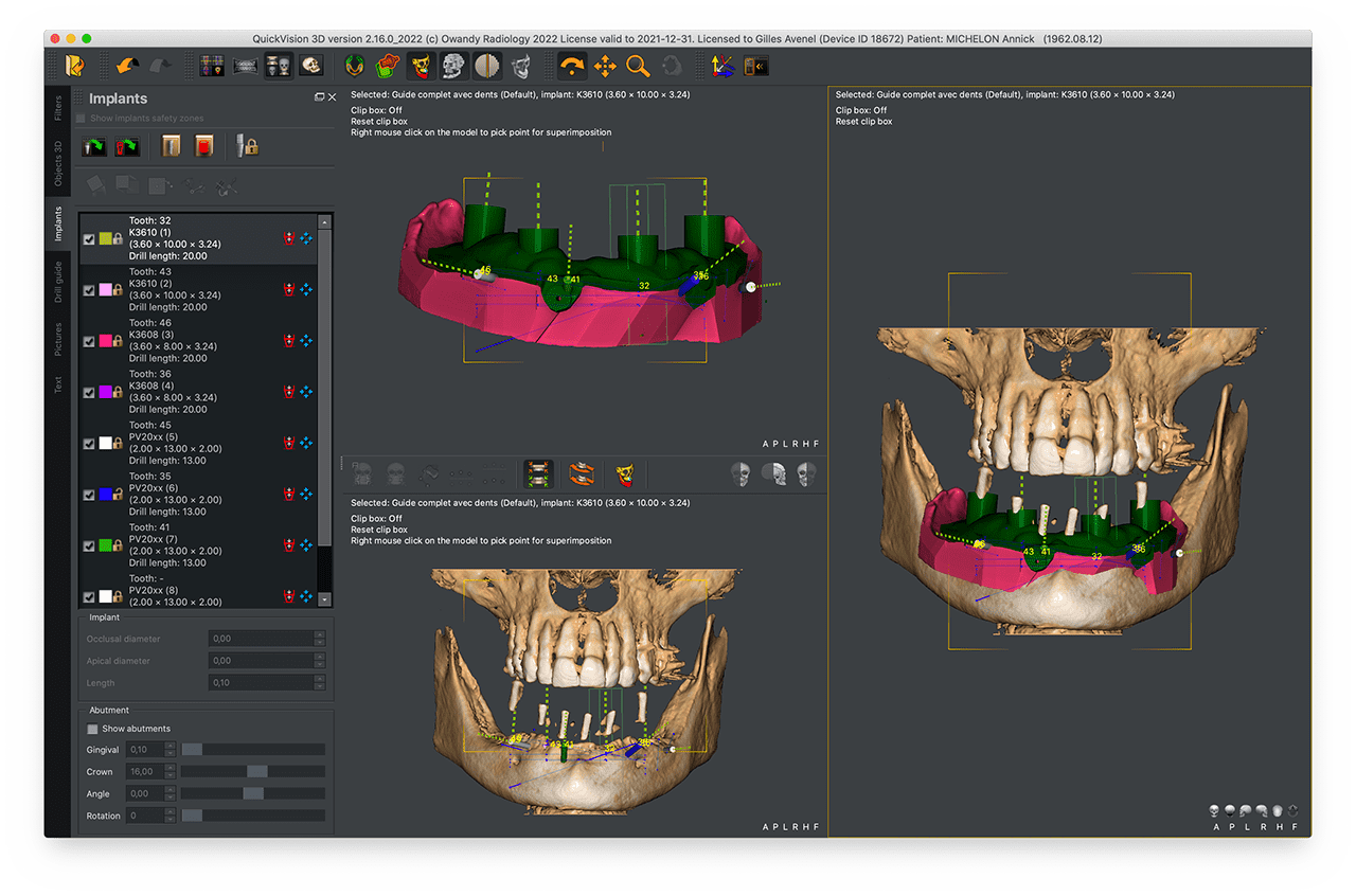
QuickVision 3D
Implant planning software
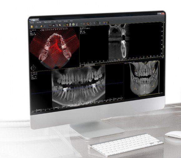
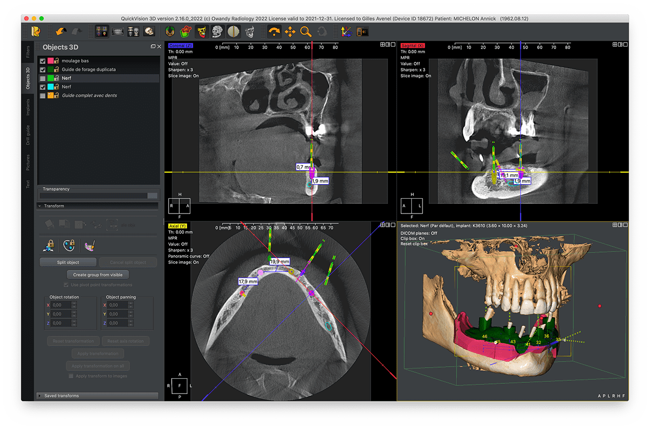
QuickVision 3D is a comprehensive software package that generates panoramic images, cross-sections and bone models based on axial image readings, enabling you to identify the mandibular canal and show the 3D bone model to calculate bone density.
QuickVision 3D allows you to simulate implant placement on 2D and 3D models. To facilitate surgery, the patient's main anatomical features are identified: the exact location of the implant, possible collisions and many other clinical aspects.
Quickvision 3D implant planning software will be your best ally in making prosthetic implant surgery faster, safer and more efficient.
Other products to discover
Discover more products and find the one that's right for you.
Panoramic
I-Max PRO

Panoramic
I-Max Ceph PRO
Cone beam
I-Max 3D Ceph XPRO
